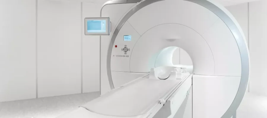
MRI in Turkey
MRI in Turkey is a widely used, non-invasive imaging method available in both public and private healthcare facilities.
Magnetic Resonance Imaging (MRI) in Turkey offers high-resolution scans for brain, spine, joints, and internal organs without radiation exposure. The procedure is safe, painless, and commonly used to diagnose neurological, musculoskeletal, and abdominal conditions. In private hospitals, MRI costs typically range from $300 to $800 depending on the body part and whether contrast is used.
What is MRI?
MRI is a radiation-free diagnostic method that uses a magnetic field to image organs and tissues in the body in detail.
Magnetic Resonance Imaging (MRI) is used to examine the brain, spine, muscles, joints, abdomen, and pelvic organs. It plays an important role in the early diagnosis of diseases by providing high-resolution images. MRI is a painless and non-invasive procedure, generally lasting 15-45 minutes.
It has no side effects except in cases where a contrast agent may be needed. It can be safely applied to pregnant women and children. Furthermore, MRI shows tumors, hernias, vascular occlusions, and internal organ disorders in detail. Not containing radiation is one of its biggest advantages.

How Does MRI Work?

MRI works with a system that creates images by stimulating hydrogen atoms in the body with a strong magnetic field and radio waves.
In Magnetic Resonance Imaging (MRI), a constant magnetic field is first applied to the body. Then, radiofrequency waves are sent to stimulate the hydrogen protons in the cells. These protons produce signals as they return to their original state, and these signals are converted into an image by a computer.
The MRI device converts these signals into detailed anatomical images, enabling the detection of diseases. It is important for the patient to remain still during the procedure for image quality. It does not contain radiation and is generally painless. MRI is one of the most effective methods for soft tissue imaging.
In Which Cases Is MRI Requested?
It is among the first choices in neurology for evaluating headaches, sudden weakness, balance disorders, vision, and consciousness changes; narrowing of brain vessels, aneurysms, or minor hemorrhages are carefully investigated. It provides detail for spine pain, suspected neck-waist hernia, and nerve root compression. In orthopedics, tears in the meniscus, anterior cruciate ligament, labrum, and cartilage damage are imaged with high accuracy.
Differentiation of masses in the liver, pancreas, kidneys, and pelvic organs; determining the extent of endometriosis or fibroids in the uterus and ovaries; and target-oriented examinations of the prostate are also performed with this method. In the cardiovascular field, myocardial viability, cardiomyopathies, and congenital vascular variations can be shown. It is also widely used in oncology for evaluating spread and post-treatment follow-up.
What to Consider Before an MRI Scan?
Preparation directly affects image quality and comfort. You should not have any metal or magnetically affected items such as watches, jewelry, hairpins, credit cards, or keys on you; you should inform us if you use a hearing aid or prosthesis. Conditions such as pacemakers, brain aneurysm clips, middle ear implants, spinal cord stimulators, or a history of metallic foreign bodies must be evaluated beforehand; although many current devices are MR-compatible, a special protocol may be required based on manufacturer information. If contrast is to be given, kidney functions and allergy history are reviewed.
Those with claustrophobia can practice with breathing and relaxation exercises; low-dose anti-anxiety medications may be planned if deemed necessary. In children, the decision for sedation for immobility during long examinations is made together with the pediatrician and anesthesia team.
How Long Does an MRI Scan Take?
The duration varies depending on the area examined and the protocol used. Scans of a single region like the brain or knee are usually short, while multi-region examinations or detailed programs for moving organs like the heart may take longer. The addition of contrast phases and the operation of special sequences increase the total time.
The duration depends not only on the device performance but also on the patient’s compliance with breathing commands and staying still during the scan; because movement can blur the image and require sequence repetition.
What is the Difference Between Open MRI and Closed MRI?
Closed systems are tunnel-form devices with high magnetic field strength; they generally provide higher resolution and shorter scanning time. Open systems, on the other hand, offer an architecture open on three sides; this increases comfort for patients with larger body sizes, severe claustrophobia, or children.
However, since the field strength is lower in some open systems, the resolution and duration equation may differ. The most suitable option is determined by evaluating the clinical necessity and the conditions the patient can tolerate; the aim is to obtain the necessary information with the highest possible quality and in the most comfortable way.
When Are MRI Results Available?
The reporting time varies according to the center’s workflow and the scope of the examination. In emergencies, critical findings are shared quickly with the physician; thus, treatment is planned without delay. In routine applications, image processing, comparison with previous examinations, and writing a report focused on the clinical question may take time.
Advanced sequences added to the protocol, such as contrast phases, diffusion, perfusion, and spectroscopy, increase the layer of interpretation. Most centers adopt an approach of first providing preliminary information, followed by the final report; thus, the patient and physician follow the process with clear steps.
Is an MRI Scan Harmful?
The method does not contain ionizing radiation; therefore, it is considered safe in most cases. Magnetic fields and radio waves do not cause known permanent damage to human tissue. However, a safety check is mandatory as the strong magnetic field may interact with some implants and devices.
In examinations using a contrast agent, special precautions may be required for individuals with kidney function impairment or, rarely, allergic reactions. Consequently, with the correct indication, appropriate patient selection, and standard protocols, the risk is quite low; the diagnostic benefit obtained significantly outweighs this risk in most scenarios.
Who Should Not Have an MRI?
Absolute prohibitions are gradually decreasing because many modern implants are manufactured to be MR-compatible. However, old-style pacemakers, some brain aneurysm clips, and metal fragments whose safety in the body has not been verified may require either postponing the examination or opting for different methods.
Severe claustrophobia, uncontrolled movement disorders, and inability to remain still during long protocols can negatively affect image quality; in these situations, a scan under anesthesia or alternative imaging methods may be considered. Postponing the procedure during the first trimester of pregnancy unless absolutely necessary is clinical practice; the decision is always made based on a risk-benefit assessment.
What is Felt During an MRI?
Rhythmic and sometimes loud noises of the device are heard throughout the scan; these sounds are the natural result of rapid currents within the magnetic field. Centers provide headphones or earplugs to reduce this noise; music may be provided in some places. A sensation of warmth or tingling in the body is rarely reported and is generally harmless. When contrast is given, a brief feeling of warmth in the arm or a metallic taste in the mouth may be felt.
The most important issue is compliance with breathing and immobility commands; a few minutes of patience makes a big difference in image quality. Individuals with claustrophobia benefit from simple methods such as closing their eyes, focusing on their breath, and maintaining communication with the technician.
MRI in Turkey Prices 2025
In 2025, MRI prices in private hospitals in Turkey generally range between 4,000 TL and 7,500 TL.
MRI (Magnetic Resonance Imaging) prices vary depending on the city where the hospital is located, the magnetic strength of the device (e.g., 3 Tesla MRI devices may have higher fees), and the number of regions to be imaged or the use of a contrast agent.
Additionally, in some cases, laboratory tests performed beforehand (e.g., kidney functions) and the need for sedation are reflected in the budget plan. The difference between public and private institutions, contract conditions, and reimbursement policies determine the patient’s share; contact us now for MRI prices.
Frequently Asked Questions
Is MRI performed on an empty stomach?
- Fasting is not mandatory for most examinations; however, eating lightly a few hours before the scan provides comfort for those prone to nausea.
- In some protocols, such as for the abdomen and bile ducts, short-term fasting may be requested not to affect organ movement and bile flow; the center’s instructions before the appointment should be followed.
- If contrast is to be administered, water consumption is usually allowed; adequate hydration facilitates intravenous access and contrast tolerance.
Are clothes removed during an MRI scan?
- Pieces containing metal such as buttons, zippers, bra wires, and similar items are removed because they can distort the image.
- Patients are provided with MR-compatible gowns in most centers; personal belongings are kept in a locked cabinet.
- Some types of heating patches, products containing ferromagnetic particles in makeup, and temporary tattoos should be reported before the scan as they may cause unwanted heating.
Is having an MRI scan painful?
The procedure is painless; what is felt is mostly the sounds of the machine and the need to remain still. A brief pinprick may be felt when the intravenous line is opened. In contrast-enhanced examinations, a warm sensation in the arm or a metallic taste in the mouth may be felt; these sensations pass quickly. For those with claustrophobia, relaxation focused on breathing and continuous communication with the technician makes the process easier.
Can music be listened to during an MRI scan?
Many centers offer a music option with headphones. This helps to tolerate the sounds of the machine and makes the duration more comfortable. However, music may be briefly lowered during some sequences due to breath-hold commands. For your safety, the headphones are provided by the center as MR-compatible equipment; personal electronic devices are not allowed inside.
