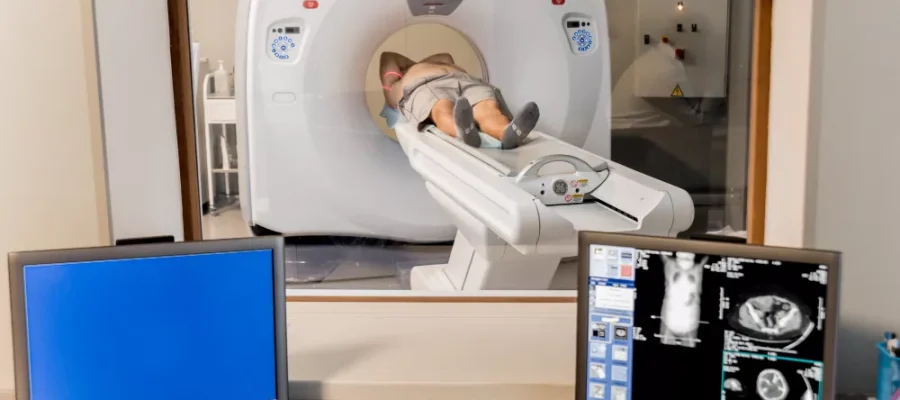
CT Thorax is a computed tomography method that provides detailed imaging of the chest cavity and lungs. It is also called chest tomography. In this method, three-dimensional images are obtained with a tomography device. Expert physicians use the resulting three-dimensional detailed cross-sections to make a diagnosis and create a treatment plan. CT Thorax is used in the diagnosis of many diseases, especially lung diseases. The increase in lung-related ailments has set the stage for the frequent use of this imaging method. The images resulting from computed chest tomography help to see the level of the disease along with its diagnosis. Recently, following the Covid-19 pandemic, the course and stage of the disease can also be clearly monitored with this imaging method, according to published guidelines.
For Which Diseases is CT Thorax Performed?
CT Thorax, which is the biggest aid to expert physicians in evaluating the organs in the chest cavity, is generally performed in case of the following health problems, although it depends on the physician’s request:
– Congenital anomalies
– Lung tumor
– Chest trauma
– Pneumonia
– Bronchitis
– COPD, emphysema
– Lung nodule follow-up
– Blockage in the lung vessels,
– Family history of lung diseases, individuals belonging to a risk group
– Aorta diseases
– Covid-19 Coronavirus disease course and follow-up
Can It Be Used in Coronavirus Disease Analysis?
Although experts underline that this scan is not a Covid-19 diagnostic method, the effect of the coronavirus process on the patient’s lungs can be monitored with computed tomography technology. For this reason, this scan may be requested from coronavirus patients in some situations to track the course of the disease. In some cases, a patient who does not show clear coronavirus symptoms may be concluded to have a suspicion of Covid-19, according to the guidelines followed by radiology specialists after performing this scan. Therefore, this computed tomography scan should not be considered an alternative to the Covid-19 diagnostic test, but rather a method used in the analysis of the treatment process.
How is the Scan Performed?
The **CT Thorax** scan is simple and fast: the patient lies on a stretcher inside a machine that is circular in shape, open at the front and back. Before lying down on the stretcher in the middle of the machine, the patient must remove any metallic or magnetic objects from their body. The patient lies supine on the stretcher, and the machine, which focuses on the chest area, performs the scan as the stretcher moves forward. The patient is asked to remain motionless during the scanning process, which lasts an average of a few seconds. The images obtained as a result of the scan are transferred to the computer and processed to analyze whether there are any risks or problems, and a report is written accordingly. As a result of the imaging, the tissue, vessel, nerve, and bone structures of the internal organs can be examined. This scan is offered at the Sonomed Imaging Center using a device with **256-Slice and low-dose technology**. The result of the imaging is instantly transferred to the computer and presented for the doctor’s review. Patients do not feel any pain or ache during the scan. It is possible to return to the daily routine after a process that takes only a few minutes.
