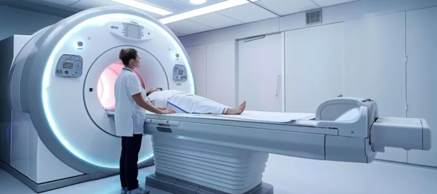
The brain is among our most sensitive organs. For this reason, the medical imaging method used to detect any problem in the brain must also be suitable for its delicate structure. Brain MRI (Magnetic Resonance Imaging) is an imaging method used to detect suspected conditions or damages that may occur in the brain. MRI, or full name magnetic resonance imaging, performs scans using radio waves. This imaging method does not contain radiation. Therefore, MRI examinations are widely requested by doctors.
Brain MRI in Turkey
A brain MRI in Turkey provides detailed, radiation-free imaging for neurological conditions in both public and private hospitals.
This non-invasive scan is used to diagnose brain tumors, strokes, multiple sclerosis, and memory-related disorders. In 2026, the cost of a brain MRI in private clinics in Turkey typically ranges from $200 to $380, depending on the location and whether contrast is required. State hospitals offer brain MRIs for free or at reduced rates under SGK coverage. Major cities like Istanbul, Ankara, and Izmir provide modern imaging centers with experienced radiologists and English-speaking staff. Brain MRI in Turkey combines affordability, accuracy, and accessibility, making it a popular choice for local and international patients.
How is a Brain MRI Scan Performed?
Sonomed Imaging Center provides brain MRI services with an expert radiology staff. The important point here is that the imaging during the MR scan is done efficiently. Since the brain is a very detailed organ, images must be analyzed with millimetric calculations, and the scan must be performed clearly. In this regard, Sonomed offers services with devices that can provide clear images and are sensitive to sound precision, utilizing advancements in MR technologies.
When performing a brain MRI scan, it is necessary to be in a lying position on the sliding table of the MRI machine. At this stage, it is important that you follow the instructions of the radiology team and remain in the lying position without moving as much as possible throughout the scanning time. The scan is completed in the MRI device while you are lying down. For those who hesitate to enter the MRI device, wider bore applications have been introduced in the devices. In this way, efforts have been made to minimize effects such as claustrophobia and fear of enclosed spaces. The duration of a brain MRI scan may vary. The Sonomed team will provide detailed information on this matter.
Points to Consider
The most important point to consider when undergoing a Brain MRI scan is not to enter the MRI device with metal objects. Since the device works like a magnet, metal objects create a hazard during the scan. All metal items such as watches, rings, glasses, and earrings must be removed. Since metal objects are not allowed in the scan, people with pacemakers cannot have a brain MRI. Also, those with implanted hearing aids cannot enter the device. The same applies to those who carry any form of metal in their body (such as shrapnel/lead). After you have made your appointment, the points you need to pay attention to before and during the examination will be communicated to you in detail.
How Long Does a Brain MRI Scan Take?
The duration varies depending on the region examined and additional sequences. A standard brain protocol is mostly completed within 15–25 minutes. The addition of contrast-enhanced phases and the planning of advanced sequences such as perfusion, spectroscopy, or tractography can prolong the duration.
Since motion artifact reduces image quality, remaining still during the scan effectively shortens the total time because it reduces the need for repeating sequences. In emergency scenarios (such as stroke, etc.), information can be obtained much faster with diffusion-weighted abbreviated protocols; in elective conditions, the scope is determined according to the target question.
What Does a Brain MRI Show?
The method strongly reveals soft tissue contrast, allowing for the selection of small hemorrhages, edema, demyelinating plaques, cortical dysplasias, microcysts, and millimetric masses. The relationship of tumors such as pituitary microadenomas, craniopharyngiomas, and meningiomas with adjacent vessel-nerve structures is clarified. The diagnostic certainty increases when lesions such as acoustic neuromas in the inner ear and brainstem, venous sinus thrombosis, or arterial stenoses are evaluated with MR angiography.
Diffusion imaging reflects cellular water movements, showing early ischemia, perfusion indicates the hemodynamic state, and spectroscopy shows metabolite profiles. This multi-layered data plays a critical role not only in diagnosis but also in monitoring treatment response and predicting prognosis.
Is Brain MRI Harmful?
Magnetic resonance does not contain X-rays or similar ionizing radiation. In this respect, its safety profile is high even in repeated controls. There is no clinical evidence that magnetic fields and radio waves cause permanent damage to human tissue. The risk is mostly related to MR-incompatible implants and rare contrast reactions.
Allergy to gadolinium-based contrast agents is a very low probability; the decision for contrast is individualized in those with impaired kidney function. Warming risks are controlled by paying attention to the specific absorption rate (SAR) limits in the protocols. With the correct indication, a trained team, and appropriate device parameters, the risk-benefit balance is significantly in favor of the benefit.
Difference Between Contrast and Non-Contrast MRI
Non-contrast protocols may be sufficient to show brain anatomy, edema, hemorrhage, ischemia, and many structural changes. However, contrast is necessary to provide clear answers to some questions. For example, tumor–scar differentiation, meningeal involvement, dynamic evaluation in pituitary microadenoma, emphasis of active inflammation, and protocols developed for vessel walls benefit from contrast. Contrast enhances the lesion borders by uptake due to increased permeability and vessel density in pathological tissues. Nevertheless, contrast is not “automatic” in every case; the decision is made based on the clinical question, previous examinations, and safety assessment.
When are Brain MRI Results Available?
The reporting time varies depending on the center’s workload and the scope of the protocols used. Preliminary information is quickly given to physicians when acute findings (acute ischemia, dangerous mass effect, etc.) are detected. Routine reports are prepared with image processing, comparison with previous examinations, and interpretation focused on the clinical question. In multi-layered examinations (with and without contrast, perfusion, spectroscopy), the evaluation time may be longer. Discussing the final report with the physician face-to-face or in an online consultation ensures the correct transfer of findings to the treatment plan.
Brain MRI in Turkey Cost 2026
In 2026, brain MRI prices in private hospitals in Turkey generally range between 6,500 TL and 15,000 TL.
Pricing varies according to the scope of the examination (non-contrast/contrast-enhanced), additional sequences (perfusion, spectroscopy, tractography), device technology, report delivery speed, and the institution’s contract–reimbursement conditions. A standard single-region scan and a multi-parametric, comparative report requested scope are not in the same budget. In necessary cases, sedation, intravenous access materials, laboratory tests (kidney functions), and consultation processes may affect the total cost. The most accurate planning is clarifying the clinical question and determining the scope accordingly; **contact us now for Brain MRI prices**.
Frequently Asked Questions
Is Brain MRI performed on an empty stomach?
- Fasting is not required for most scans; coming with light nourishment provides comfort.
- The center may recommend short-term fasting if the abdomen-pelvis is planned simultaneously or if sedation is considered.
- If contrast is to be administered, drinking water is generally allowed; good hydration facilitates the placement of an intravenous line.
- Follow your physician’s instructions for diabetes medications and regular treatments; time adjustments are made in some cases.
What is felt during a Brain MRI scan?
The examination is painless. You lie on the table, and while inside the coil placed around your head, you hear the rhythmic, sometimes loud noises of the machine; this is tolerated with earplugs. Remaining still is your most important “task”; closing your eyes and focusing on your breath shortens the time. If contrast is administered, a brief warmth in the arm or a metallic taste in the mouth may be felt; this disappears within seconds. Those with claustrophobia concerns can relax by talking to the technician; the device has an emergency button and visual-audio communication.
Does Brain MRI show headaches?
The headache itself cannot be visualized; however, structural causes that may lead to headaches can be detected or ruled out. Sinus problems, vascular malformations, masses, signs of hemorrhage-ischemia, and indirect clues indicating pressure changes are evaluated within this scope. The examination is mostly normal in primary headaches (such as migraine, tension-type, etc.); this provides reassurance that there is no dangerous cause and allows the treatment to be planned on a clinical basis.
Is Contrast-Enhanced Brain MRI clearer?
- **Lesion boundary and activity:** Contrast sharpens the distinction between tumor–inflammation–scar by emphasizing the permeability of the pathological tissue.
- **Meninges and nerve pathways:** Selectivity increases in meningeal involvement, nerve root–ganglion pathologies, and vascular inflammation.
- **Is it mandatory for every case?** No. Non-contrast protocols may be sufficient in the early stage of ischemia, traumatic hemorrhages, or typical demyelinating plaque follow-ups.
- **Safety:** Gadolinium reaction is rare; the decision is made based on kidney functions, and the dose and indication are individualized.
Can normal life be resumed after a Brain MRI?
Generally, yes. If sedation was not administered, you can return to daily activities immediately after the scan. If contrast was received, drinking plenty of water during the day provides comfort. If unusual symptoms such as rash, shortness of breath, or dizziness develop, it is advisable to contact the health team; these symptoms are not common and are usually controlled quickly.
